Oral Medicine & Radiology
The Department of Oral Medicine and Radiology deals with the diagnosis and medical management of oro facial diseases and the oral manifestations of systemic disorders. It also deals with the maxillofacial imaging and dental management of medically compromised patients (patients with medical problems).
Oral Medicine Diagnosis & Radiology is the first department where the patient is received; elaborate history is taken with thorough examination and investigations that aid in arriving at a diagnosis are done. Patients are medically managed with required alterations in medications and other medical advises are given.
Radiographic Diagnostic Tools :
- Completely Digital Radiography
- Orthopantamogram (Opg)
- Rvg
- Bite Wing Radiograph
- Cephalometric Radiograph
Treatments
Dental Caries
Dental caries, also known as tooth decay or a cavity, is an infection, bacterial in origin, that causes demineralization and destruction of the hard tissues of the teeth (enamel, dentin and cementum). It is a result of the production of acid by bacterial fermentation of food debris accumulated on the tooth surface.If demineralization exceeds saliva and other remineralization factors such as from calcium and fluoridated toothpastes, these once hard tissues progressively break down, producing dental caries (cavities, holes in the teeth). Today, caries remain one of the most common diseases throughout the world. Cariology is the study of dental caries. Depending on the extent of tooth destruction, various treatments can be used to restore teeth to proper form, function, and aesthetics, but there is no known method to regenerate large amounts of tooth structure. Instead, dental health organizations advocate preventive and prophylactic measures, such as regular oral hygiene and dietary modifications, to avoid dental caries.
Grossly Decayed Teeth
This patient presented as an emergency with pain in the upper left quadrant. The radiograph shows the problem. There is a large carious lesion present and invading the nerve. The patient chose to have the tooth extracted. Extensive work could have been done to save it, but the end result would leave you with a compromised tooth.
Dental Abscess
A dental abscess (also termed a dentoalveolar abscess, tooth abscess or root abscess), is a localized collection of pus associated with a tooth. The most common type of dental abscess is a periapical abscess, and the second most common is a periodontal abscess. In a periapical abscess, usually the origin is a bacterial infection that has accumulated in the soft, often dead, pulp of the tooth. This can be caused by tooth decay, broken teeth or extensive periodontal disease (or combinations of these factors). A failed root canal treatment may also create a similar abscess.
Periapical Lesions
Periapical lesions often develop slowly and do not become very large. Patients do not experience pain unless there is acute inflammatory exacerbation. These lesions are often diagnosed during routine radiographic exams. Some periapical lesions become large and, in cases of large radiolucencies, they may be diagnosed in the absence of any patient complaint. Sometimes, symptoms such as mild sensitivity, swelling, tooth mobility and displacement may be observed in these cases. Large periapical lesions are often associated with anterior maxillary teeth, probably due to traumatic injuries. These lesions could be classified as granulomas, pocket cysts (also called bay cysts) and true cysts. Granulomas are usually composed of solid soft tissue, while cysts have a semi-solid or liquefied central area usually surrounded by epithelium. Pocket cysts have an epithelial lining that is connected with the root canal, and true cysts are completely lined with epithelium and not connected with the root canal.
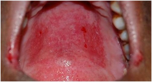
ulcers
An ulcer is a discontinuity or break in a bodily membrane that impedes the organ of which that membrane is a part from continuing its normal functions.
Leukoplakia
Leukoplakia is a condition where areas of keratosis appear as adherent white patches on the mucous membranes of the oral cavity. Leukoplakia may affect other gastrointestinal tract mucosal sites, or mucosal surfaces of the urinary tract and genitals.
Oral cancer
Oral cancer or mouth cancer, a subtype of head and neck cancer, is any cancerous tissue growth located in the oral cavity.It may arise as a primary lesion originating in any of the oral tissues, by metastasis from a distant site of origin, or by extension from a neighboring anatomic structure, such as the nasal cavity. Alternatively, the oral cancers may originate in any of the tissues of the mouth, and may be of varied histologic types: teratoma, adenocarcinoma derived from a major or minor salivary gland, lymphoma from tonsillar or other lymphoid tissue, or melanoma from the pigment-producing cells of the oral mucosa.
Oral Submucous Fibrosis
Oral submucous fibrosis (OSF) is a chronic, complex, irreversible, highly potent pre-cancerous condition characterized by juxta-epithelial inflammatory reaction and progressive fibrosis of the submucosal tissues (lamina propria and deeper connective tissues). As the disease progresses, the jaws become rigid to the point that the sufferer is unable to open his mouth.The condition is linked to oral cancers and is associated with areca nut chewing, the main component of betel quid. Areca nut or betel quid chewing, a habit similar to tobacco chewing, is practiced predominately in Southeast Asia and India, dating back thousands of years
Lichen planus
Lichen planus is an inflammatory skin condition, characterized by an itchy, non-infectious rash of small, polygonal (many sided) flat-topped pink or purple lesions (bumps) on the arms and legs. Other parts of the body may also be affected, including the mouth, nails, scalp, vulva, vagina, and penis. Involvement in the scalp can result in hair loss - sometimes permanent.
